41 labeled histology slides
Free templates about anatomy for Google Slides & PPT Human anatomy is the study of the structure of the human body and its components. It includes a detailed examination of all parts of the body, from the smallest cell to the largest organ systems. The field of anatomy seeks to understand how each component works in harmony with others... Multi-purpose. 16:9. Slides of Histology | Anatomy and Physiology I - Lumen Learning Slides of Histology Learning Objectives Be able to describe the functions of cells commonly found in connective tissue and identify them. Be able to recognize interstitial (fibrillar) collagens and elastic fibers at the light and electron microscopic levels.
Uniform Labeling of Blocks and Slides in Surgical Pathology The panel addressed the overarching question, "What are the essential elements for the proper labeling of paraffin blocks and microscopic slides in the routine practice of surgical pathology?" and developed 12 recommendations, including: Laboratories should ensure that all blocks and slides are clearly labeled using two patient identifiers.

Labeled histology slides
1st Year MBBS Histology Slides and Identification Points (FINAL).pdf 1st Year MBBS Histology Slides and Identification Points (FINAL).pdf - Google Drive. Histology Slides - Microscope.com As a counterpart to gross anatomy, histology is an invaluable method for learning biology. This excellent collection of 50 slides taken from human tissues is an excellent medical histology resource for instruction at the high school, college and graduate levels. Most organ systems and tissues are represented. 110 Simple columnar epithelium. HistologySlide - Identification of Microscope Slides Histology Slide Identification with Identifying Characteristics Collection of microscope images, labeled diagrams, videos, and more Latest from HistologySlide Slide Identifying Points Slide Identifying Points Slide Identifying Points Slide Identifying Points Slide Identifying Points Slide Identifying Points Slide Identifying Points
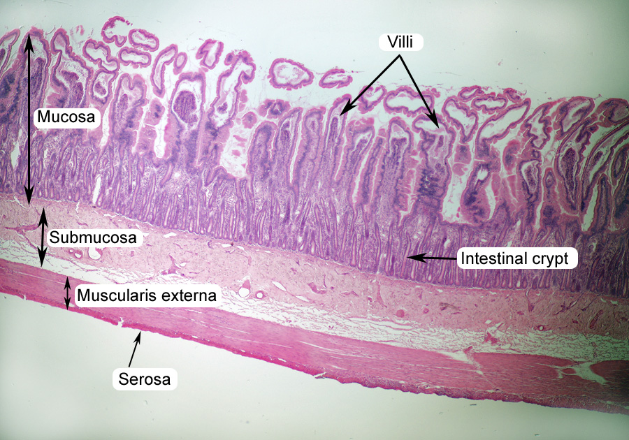
Labeled histology slides. Learn histology faster With quizzes and flashcards | Kenhub With Kenhub's huge library of histology slides, of course! In our histology atlas, we clearly highlight a given structure on our slides. Comparing several of these slides next to one another is a great way to get a feel for how one tissue differs from another. Enter: our labeled and unlabeled histology tissue identification quiz worksheets. Online Histology Made Easy Slides Atlas - MedicForYou W e have prepared an online atlas of histology that has the following histology slides terming it as Histology made easy. These histology slides can be used for practical exams in First Year MBBS while others may find it useful in their ways. Hope the following slides help you in exams and learning it better, though you will have to refer to a histology atlas book. Ileum Histology Slide with Labeled Diagram and Identification Points Ileum Histology Slide with Labeled Diagram and Identification Points 04/11/2022 05/11/2021 by anatomylearner The ileum histology slide consists of the four layers like tunica mucosa, submucosa, muscular, and serosa. Here, I will show you the detailed histological features of the wall of the ileum slide with a labeled diagram. Colon Histology Slide with Labeled Diagram - AnatomyLearner Colon histology layers There are four different tunica layers in the wall of a colon microscope slide - mucosa, submucosa, muscular, and serosa. The structure of the four different layers of the colon microscope slide is almost similar to the structure of a tubular organ. Colon histology slide layers labeled diagram
all histology slide identification tricks | how to identify histology ... all histology slide identification tricks | how to identify histology slide | easy histology vivaFor Buy Anatomy Module Go Through My Website ... Histology: Labelled Slides | SchoolWorkHelper Histology: Labelled Slides Aorta Basophils Cardiac Muscle Cardiac Muscle Longtudinal Cerebellar Cortex Cerebral Cortex Spinal Cord Eosinophils Epiphyseal plate Femoral Artery Femoral Vein Howship's lacunae Jejunum lamina propria Liver Lymph node Lymphocyte Monocyte Spinal cord (silver) Neutrophil Pacinian corpuscle Peripheral Nerve Mislabeling of Cases, Specimens, Blocks, and Slides: A College of ... Mislabeled Slide.—Histologic slide was labeled with the wrong specimen/patient identification, sequence number, or letter. ... 5 histology technicians, 2 clerks, and 2 "other" workers. Table 5 lists a number of laboratory practice characteristics related to accessioning and processing in the laboratory. Gross examination and section ... Histology guide: Definition and slides | Kenhub At a histological level, both the heart and blood vessels consist of three layers: Endothelial layer - epithelial tissue formed by simple squamous (endothelial) cells. In the heart, this layer is referred to as endocardium. Muscular layer - smooth muscle in the blood vessels, cardiac muscle (myocardium) in the heart.
Histology Slide Identification Tricks | All Histology Slides In 15 ... Medico Darshil Histology Slides Identification Here in this video i tried to describe all histology Slides in 15 minutes. Identifying Epithelium | Review and Practice Questions Anatomy... Histology Microscope Slides | Carolina.com Comprehensive Medical Histology Slide Set Item #311992 $848.00 Connective Tissue Types Microscope Slide Set Item #312034 $88.00 Digestive Tract Microscope Slide Set Item #312106 $65.00 Discovering Epithelial Tissues Self-Study Unit, Microscope Slide Set Item #312008 $91.00 Epithelium Types Microscope Slide Set Item #312016 $73.00 histology human anatomy tissue slides - Quizlet Histology: Tissue slides simple squamous epithelium-oral smear Simple squamous epithelium-human lung Simple cuboidal epithelium Nucleus and cell Air sac and nucleus Simple cuboidal cell, basement membrane and connective tissue 21 Terms WhitneyGosvener tissue histology slides hyaline cartilage stratified squamous epithelium compact bone 21 Terms Histology Slides at Thomas Scientific High Quality prepared microscope slides for botany, zoology, histology, parasitology, pathology. The Histology Set of 8 slides includes: S99217 Cardiac Striated Miscle L.S., S99067 Skeletal Muscle L.S., S99040 Compact Bone, S99207 Lung Section, S99061 Stomach Fundic Region, S99032 Blood Smear…. Compare this item.

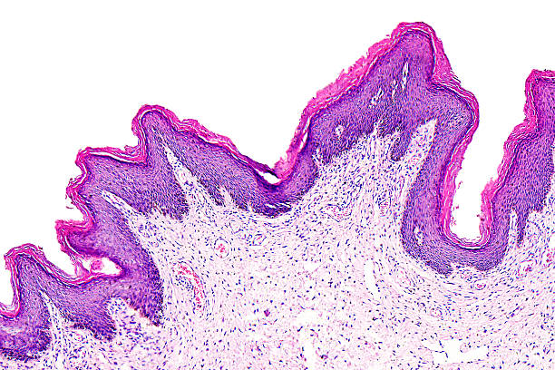
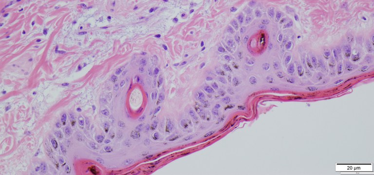
HISTOLOGY, Epithlium Lab, Scalp slide
How to examine histology slides: Techniques and tips | Kenhub How to examine histology slides. 1. Inspection: Inspect the slide using just your eyes and a good light source to first determine the shape of the prepared section. Occasionally, a specific section has a characteristic shape and is much easier to identify. e.g on the cross section of tracheal cartilage an annular preparation can be seen. 2.

taste buds slide labeled papillae taste pores - Google Search ...
Histology Slides 1 - Loyola University Chicago Welcome to the LUMEN Histology Slide Series. LUMEN
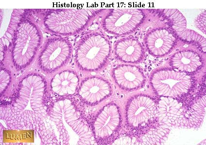
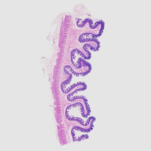
Jejunum
Virtual histopathology slide box - Knowledge @ AMBOSS The virtual histopathology slide box provides an introduction to the histology of diseased cells and tissues. Each specimen is accompanied by a caption that provides information on staining, magnification, and the structures shown. Virtual microscopy is provided in cooperation with Smart Zoom®.
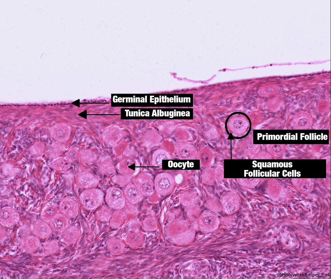
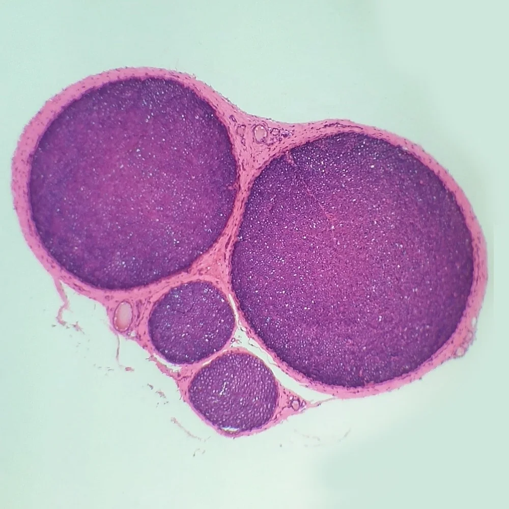
ovary-slide-labelled-histology | SchoolWorkHelper
Histology Slide Labeling & Preparation - General Data Company, Inc. Our slide labeling solutions are able to withstand the harsh chemicals, reagents and stains of a histology lab's slide staining protocols and process. Each slide is permanently identified and barcodes remain scannable before and after the staining process, as well as through review and archiving. Error-Proof Your Lab's Processes
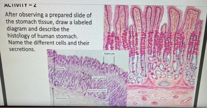
Solved ACTIVITY - Z After observing a prepared slide of the ...
Histology Guide - virtual microscopy laboratory Histology is the study of the microanatomy of cells, tissues, and organs as seen through a microscope. It examines the correlation between structure and function. Histology Guideteaches the visual art of recognizing the structure of cells and tissues and understanding how this is determined by their function.

Examples of histology slides; (left) desired cooled zone ...
Histology Slides Identification from Different Organ Systems This article will show you histology slides from the following different organs system of an animal's body with identifying features. #1. Histology slide of epithelial tissue #2. General connective tissue histology slide #3. Histology slides of special connective tissue (blood, bone, and cartilage) #4. Muscular tissue histology slide #5.

Images from histology slide preparations from specimen EG 9 ...
Small and Large Intestine | histology - University of Michigan The histology of the wall of the small intestine differs somewhat in the duodenum, jejunum, and ileum, but the changes occur gradually from one end of the intestine to the other. 1. Duodenum. Slide 162 40x (pyloro-duodenal junct, H&E) View Virtual Slide. Slide 161 40x (pylorus, duodenum, pancreas, H&E) View Virtual Slide. Look at slide 162 first.
4,500+ Histology Slides Stock Photos, Pictures & Royalty-Free ...
Types, Location, Examples and Histology Slides - AnatomyLearner If you need more epithelial tissue drawing or real slide pictures, then let me know. You may also follow anatomy learner on social media for more epithelial pictures. Don't forget to read the other articles from anatomylearner - #1. Identifying characteristics of loose connective tissue from slide pictures. #2.

Oral Histology Slide - LIP at Rs 800/piece ...
Microscope Slides of Cells and Tissues | Histology Guide This virtual slide box contains 275 microscope slides for the learning histology. Fig 023 Types of Tissue Cells and Tissues Tissues are classified into four basic types: epithelium, connective tissue (includes cartilage, bone and blood), muscle, and nervous tissue. Chapter 1 The Cell Chapter 2 Epithelium Chapter 3 Connective Tissue Chapter 4 Muscle
Histology — Department of Veterinary and Biomedical Sciences
Artery Histology - Elastic and Muscular Arteries Slides - AnatomyLearner Muscular artery histology slide labeled The tunica intima is comparatively thin than elastic arteries and consists of endothelium with a thin subendothelial layer. The subendothelial layer of the muscular artery contains collagen and elastic fibers. You may also find few fibroblasts and smooth muscle cells in this layer of the muscular artery.
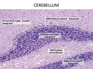
Histology of cerebrum and cerebellum
Virtual Slide List | histology - University of Michigan Resources in the University of Michigan Histology Dropbox This collection was originally compiled by Kent Christensen, Ph.D., J. Matthew Velkey, Ph.D., Lloyd M. Stoolman, M.D., Laura Hessler, and Diedra Mosley-Brower. Currently, it is curated by Michael Hortsch, Ph.D.

Histología, Veterinaria y zootecnia, Anatomía
HistologySlide - Identification of Microscope Slides Histology Slide Identification with Identifying Characteristics Collection of microscope images, labeled diagrams, videos, and more Latest from HistologySlide Slide Identifying Points Slide Identifying Points Slide Identifying Points Slide Identifying Points Slide Identifying Points Slide Identifying Points Slide Identifying Points
Histology Slides 1
Histology Slides - Microscope.com As a counterpart to gross anatomy, histology is an invaluable method for learning biology. This excellent collection of 50 slides taken from human tissues is an excellent medical histology resource for instruction at the high school, college and graduate levels. Most organ systems and tissues are represented. 110 Simple columnar epithelium.

Odontoblast - Wikipedia
1st Year MBBS Histology Slides and Identification Points (FINAL).pdf 1st Year MBBS Histology Slides and Identification Points (FINAL).pdf - Google Drive.
![PDF] Use of Labeled Histology Images with Key Identification ...](https://d3i71xaburhd42.cloudfront.net/9e03ebd5b9681a62269a4d60cd6b0af01715e6ca/2-Figure1-1.png)
PDF] Use of Labeled Histology Images with Key Identification ...
Biology W2501 :: Contemporary Biology Lab -- Histology
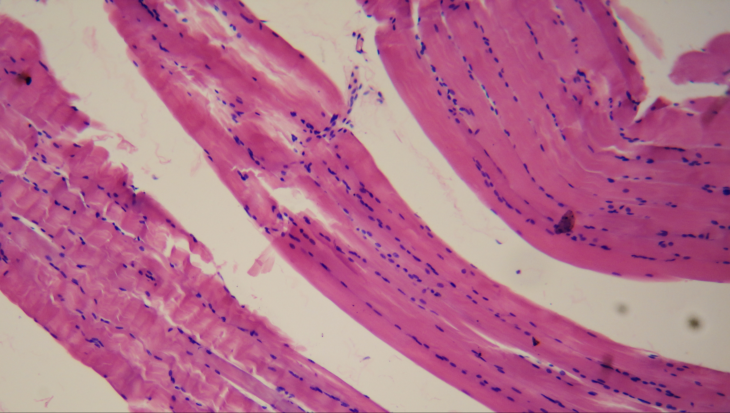
File:Smooth muscle (histology slide).jpg - Wikimedia Commons
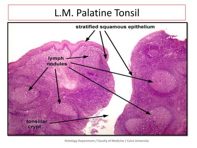
Lymphatic - Prac. Histology

Histology Slides For MBBS 1st Year [With Identification ...

Rectangular Prepared Microscope Slide Oral Histology Slides ...
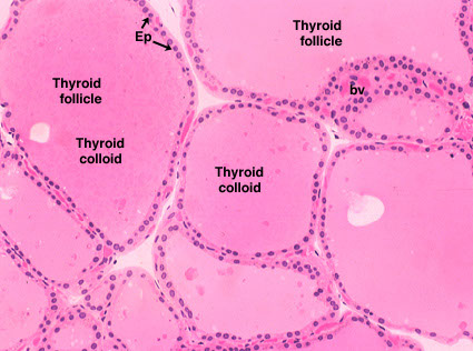
Thyroid Slide

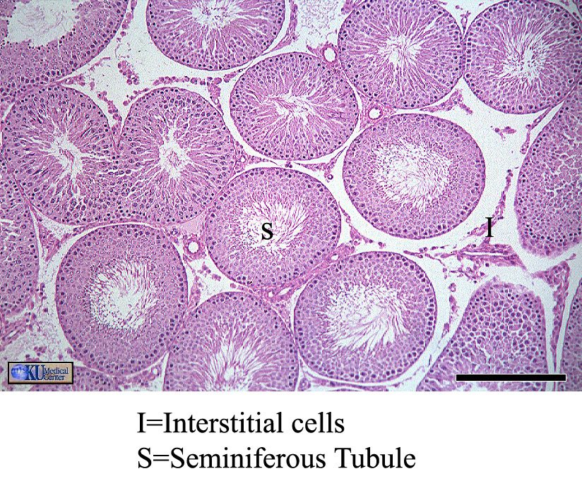
Histology of Reproductive Organs – David Fankhauser
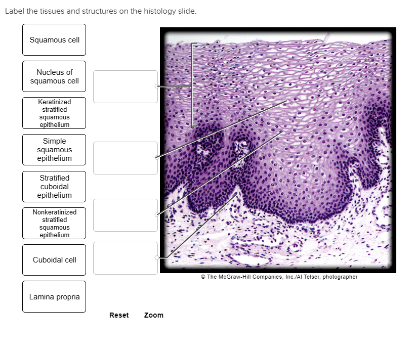
Solved Label the tissues and structures on the histology ...
4,500+ Histology Slides Stock Photos, Pictures & Royalty-Free ...
Human ileum cross-section histology slides, 7 µm sec., H&E ...
Histology Slide Jaringan Sistem Saraf Melihat Struktur Sel - Buy Histologi Slides,Slide Mikroskop,Struktur Sel Product on Alibaba.com
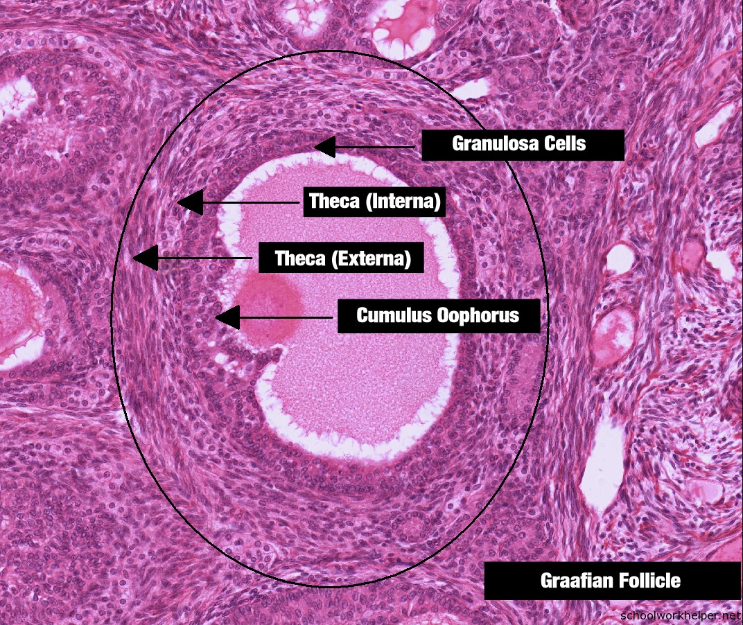
ovary-2-slide-labelled-histology | SchoolWorkHelper
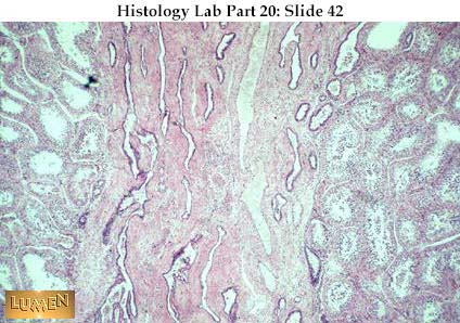
Histology Slides 1
Normal Human Histology Slide Set | Flinn Scientific

stomach histology labeled | Tissue biology, Medical ...

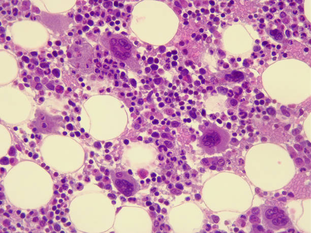
Histology slides of normal human bone marrow at 2 different ...
Larynx Histology - Epiglottis (labels) - histology slide -
Histology Slides at Thomas Scientific

Histology Slides Database: rectum histology slides

Colon histology HD wallpaper | Pxfuel

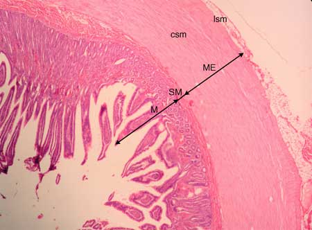
trachea-slide-labelled-histology | SchoolWorkHelper

Histology Slides Digestive, Urinary, and Reproductive A&P 102 ...

Teaching medical histology at the University of South ...
Human anatomy and physiology, Histology slides, Medical ...
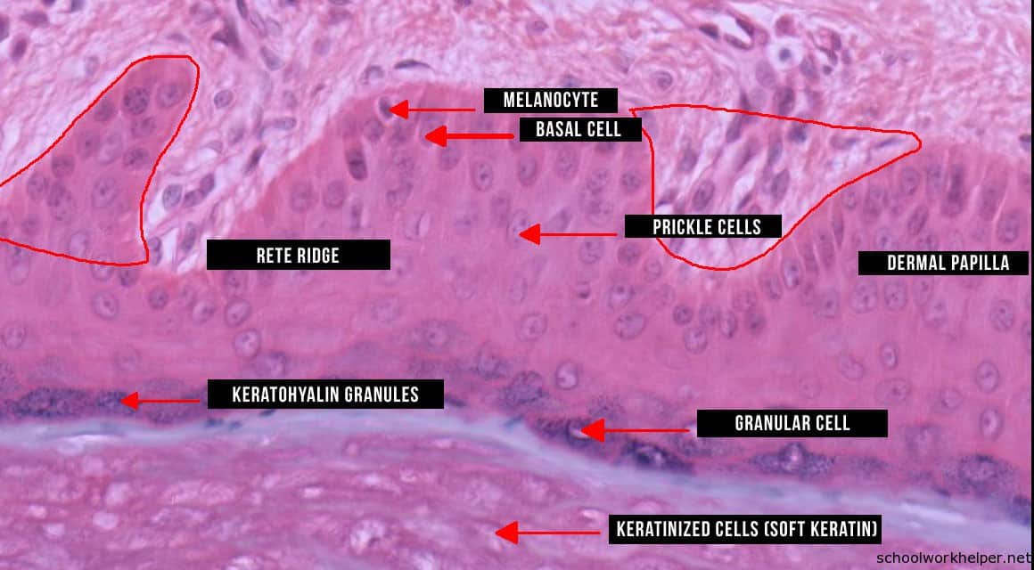
epidermis-2-slide-labelled-histology | SchoolWorkHelper
50pcs Basic human histology slides set - Xinxiang Happy ...


Komentar
Posting Komentar