40 neuron diagram labeled
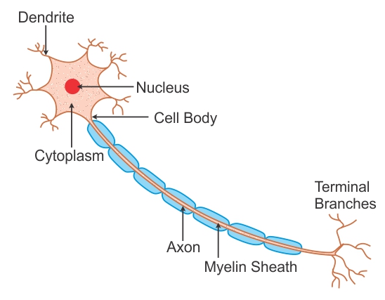
Draw neat labelled diagram of neuron. - Toppr Click here to get an answer to your question ✍️ Draw neat labelled diagram of neuron. Draw a labelled diagram of the neuron and describe the structure of ... The structure of neuron: Nerve cells or neurons are the structural and functional units of the nervous system. It consists of three major parts namely, ...
Changes to the Nutrition Facts Label | FDA Web7 mars 2022 · FDA finalized the new Nutrition Facts label for packaged foods to reflect new scientific information. It will make it easier for consumers to make better informed food choices.
Neuron diagram labeled
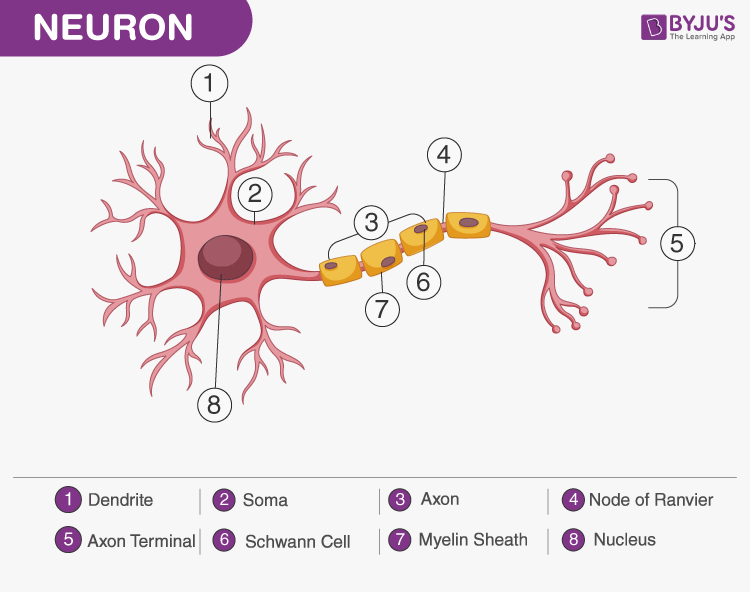
Labeled Neuron Diagram - Science Trends Types of Neurons · Unipolar: Contains only one branching body · Bipolar: Has 1 axon and 1 dendron · Multipolar: 1 axon and 2 or more dendrites · Anaxonic: Axon ... Animal Cell- Definition, Structure, Parts, Functions, Labeled Diagram Web17 sept. 2022 · Animal cell size and shape. Animal cells come in all kinds of shapes and sizes, with their size ranging from a few millimeters to micrometers. The largest animal cell is the ostrich egg which has a 5-inch diameter, weighing about 1.2-1.4 kg and the smallest animal cells are neurons of about 100 microns in diameter. Diagram Quiz on Neuron Structure and Function (Labeling Quiz) 2. In the figure, labeled ‘1’ receives impulses from adjacent neuron. It is called the Dendron Dendrite Axon Axonite. 3. In the figure, labeled ‘2’ is the short filaments from the cell body that carries impulses from dendrites to the cell body which is the Schwann cell Axonite Axon Dendron. 4.
Neuron diagram labeled. What Is a Neuron? Diagrams, Types, Function, and More - Healthline Jul 20, 2018 ... An Easy Guide to Neuron Anatomy with Diagrams ... Neurons, also known as nerve cells, send and receive signals from your brain. While neurons have ... how to draw structure of neuron/neuron diagram labelled ... - YouTube Mar 7, 2019 ... Please watch: "cell structure and functions / animal cell vs plant cell / parts of cell / ch 8 science class 8 cbse" ... Join LiveJournal WebPassword requirements: 6 to 30 characters long; ASCII characters only (characters found on a standard US keyboard); must contain at least 4 different symbols; Animal Cell- Definition, Structure, Parts, Functions, Labeled ... Sep 17, 2022 · Animal cell size and shape. Animal cells come in all kinds of shapes and sizes, with their size ranging from a few millimeters to micrometers. The largest animal cell is the ostrich egg which has a 5-inch diameter, weighing about 1.2-1.4 kg and the smallest animal cells are neurons of about 100 microns in diameter.
Unsupervised Feature Learning and Deep Learning Tutorial WebConsider a supervised learning problem where we have access to labeled training examples (x^{(i)}, y^{(i)}).Neural networks give a way of defining a complex, non-linear form of hypotheses h_{W,b}(x), with parameters W,b that we can fit to our data.. To describe neural networks, we will begin by describing the simplest possible neural network, one … Diagram Quiz on Neuron Structure and Function (Labeling Quiz) WebThis labelled diagram quiz on Neuron is designed to assess your basic knowledge in Structure and Function of Neuron. ... The part labeled ‘8’ are branches of axon that carries impulses to the synaptic knob Schwann cell glial cell Axonite neurons. 10. It is the tip of axonite that contains vesicles which releases neurotransmitters. The part labeled ‘9’ is … What is Perceptron? A Beginners Guide [Updated] | Simplilearn Web9 déc. 2022 · Every neuron holds an internal state called activation signal; Each connection link carries information about the input signal; Every neuron is connected to another neuron via connection link; In the next section, let us talk about perceptrons. Perceptron Perceptron was introduced by Frank Rosenblatt in 1957. He proposed a Perceptron learning ... Machine Learning Glossary | Google Developers Nov 7, 2022 · In that case, the neuron calculates the sigmoid of -2.0, which is approximately 0.12. Therefore, the neuron passes 0.12 (rather than -2.0) to the next layer in the neural network. The following figure illustrates the relevant part of the process:
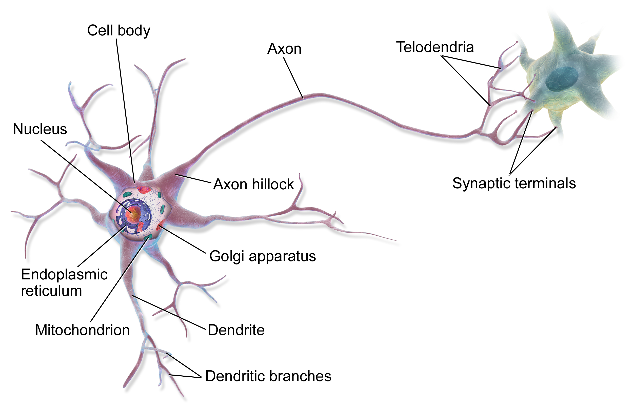
Neuromuscular Junction - The Definitive Guide | Biology ... Apr 17, 2021 · To better understand what a neuromuscular junction is, we should first look at the structure of the motor neuron terminal (1, 3), the synaptic cleft (2), and muscle membrane (4, 5). The labeled neuromuscular junction below shows these main components. Important parts of the NMJ. Motor Neuron Machine Learning Glossary | Google Developers Web7 nov. 2022 · In that case, the neuron calculates the sigmoid of -2.0, which is approximately 0.12. Therefore, the neuron passes 0.12 (rather than -2.0) to the next layer in the neural network. The following figure illustrates the relevant part of the process: active learning. A training approach in which the algorithm chooses some of the data it learns from. Active … File:Complete neuron cell diagram en.svg - Wikipedia WebEnglish: Complete neuron cell diagram. Neurons (also known as neurones and nerve cells) are electrically excitable cells in the nervous system that process and transmit information. In vertebrate animals, neurons are the core components of the brain, spinal cord and peripheral nerves. Date: 12 July 2007: Source: Own work. Image renamed from … Neuron - The Definitive Guide | Biology Dictionary Web28 juin 2020 · A bipolar neuron has the same structures as all neurons but is arranged differently. The central cell body makes a border between the dendrite end and the axon and terminal end. Dendrites are multiple and sometimes it is difficult to know which end is which in a simplified bipolar neuron diagram. Usually, the rounded shape of the terminal ...
Ultrastructure of Nerves - Classification - Neurones ...
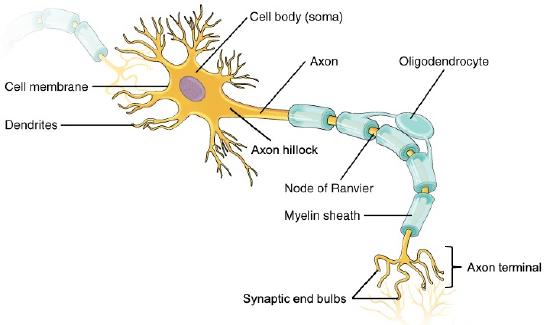
Nervous System - Label the Neuron Nervous System - Neuron: Nerve Cell. Name: Choose the correct names for the parts of the neuron. neuron, (1). axon, cell body, dendrites, nucleus ...

Simple Neuron Diagram 2d Labeled Nerve Stock Vector (Royalty ...
Simple Squamous Epithelium under a Microscope with a Labeled ... Mar 4, 2022 · Here, in the epidermis of skin labeled diagram, I tried to show you the keratinization over the cell surface. Again, the diagram also shows the cells of the different layers of skin’s epidermis. You will also find the keratinized stratified squamous epithelium on the oral cavity of an animal. Pseudostratified columnar epithelium under a ...

Diagram Quiz on Neuron Structure and Function (Labeling Quiz)
Changes to the Nutrition Facts Label | FDA - U.S. Food and ... Mar 7, 2022 · So for packages that are between one and two servings, such as a 20 ounce soda or a 15-ounce can of soup, the calories and other nutrients are required to be labeled as one serving because people ...
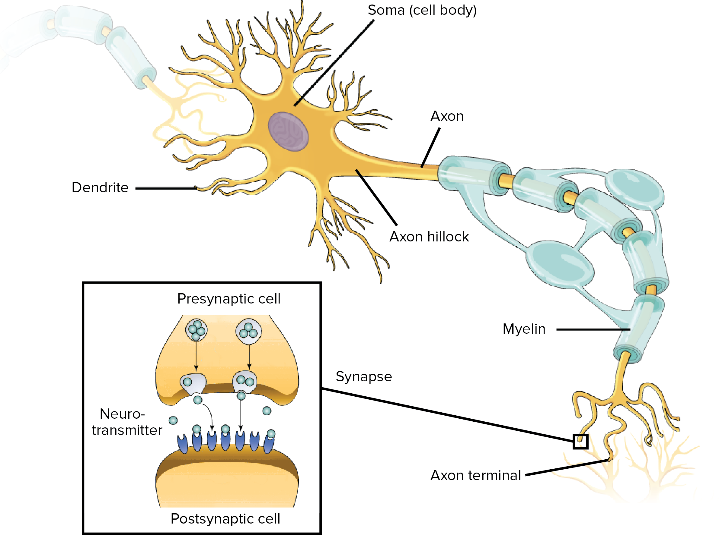
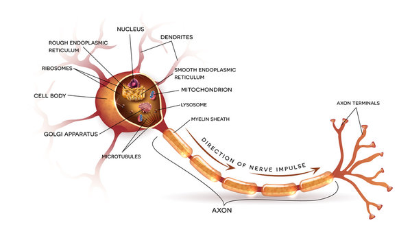
Overview of neuron structure and function (article) | Khan ...
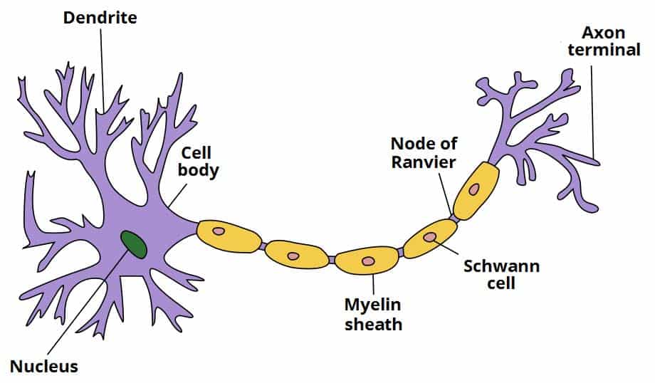
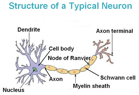
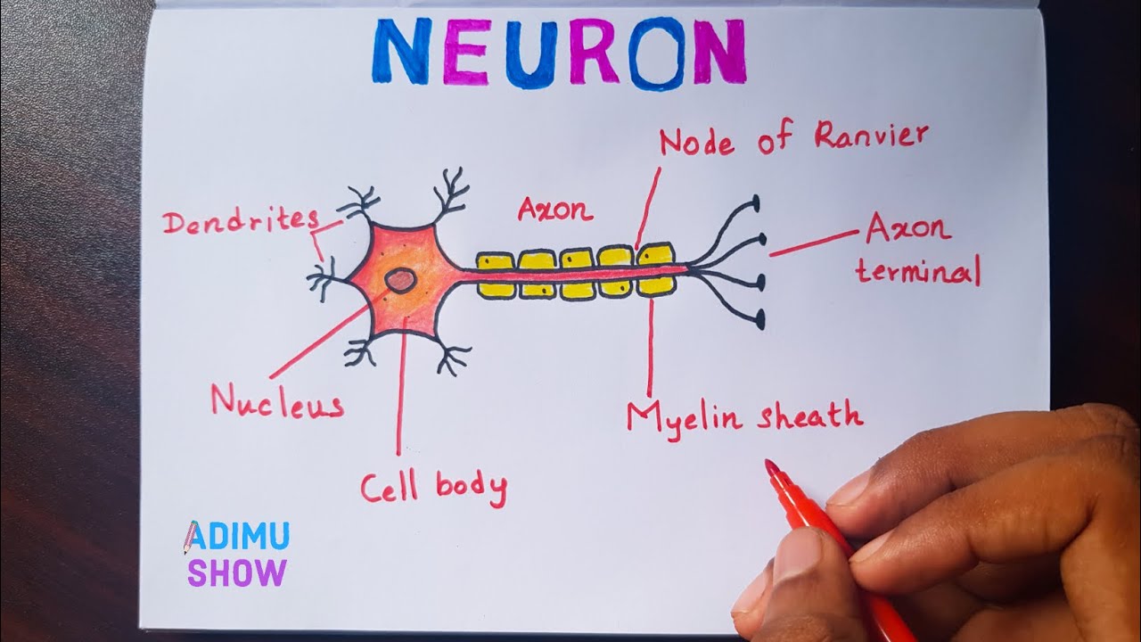
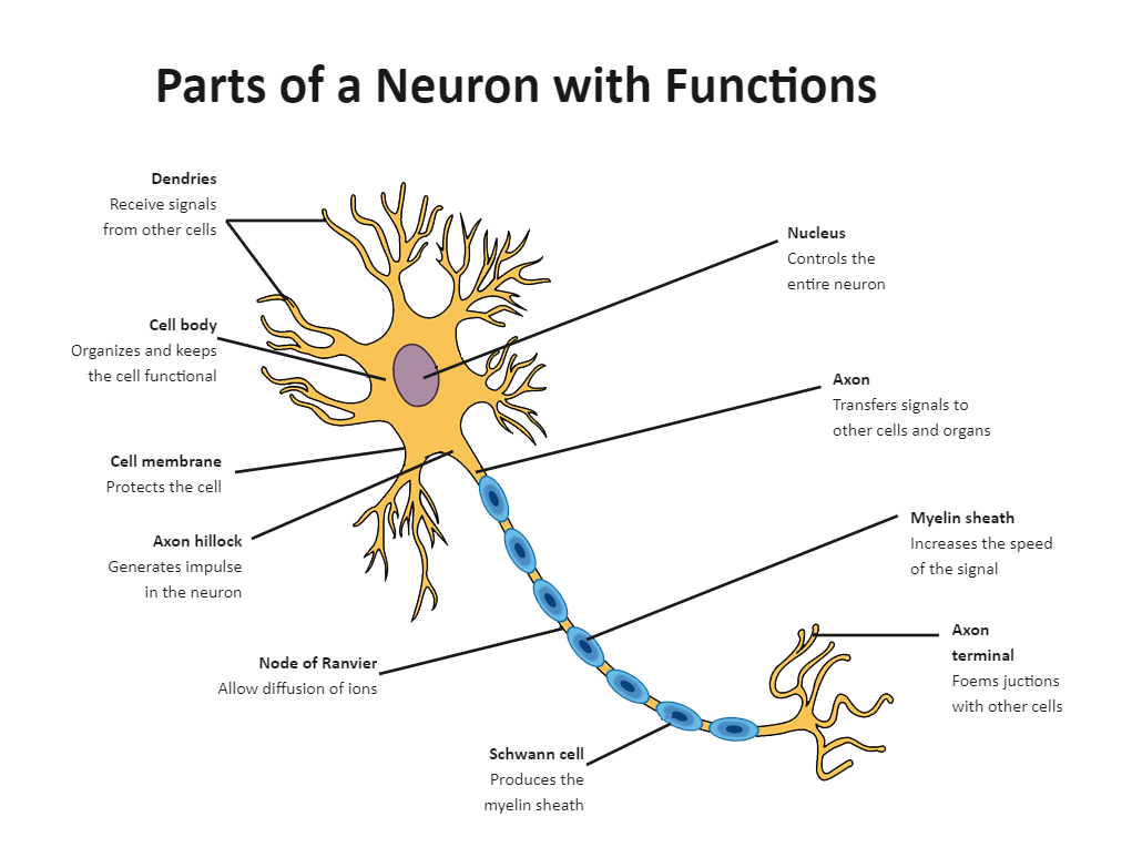
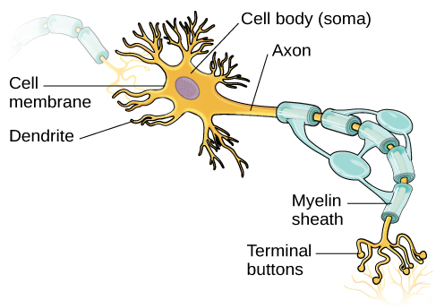
A Labelled Diagram Of Neuron with Detailed Explanations - Byju's May 27, 2022 ... Diagram Of Neuron with Labels · Dendrites–A branch-like structure that functions by receiving messages from other neurons and allow the ...
11.3: Neurons - Biology LibreTexts
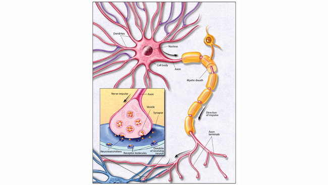
Labeled Diagram of the Neuron Stock Vector - Dreamstime.com Illustration about Labeled diagram of the neuron, nerve cell that is the main part of the nervous system. Illustration of anatomical, peripheral, ...

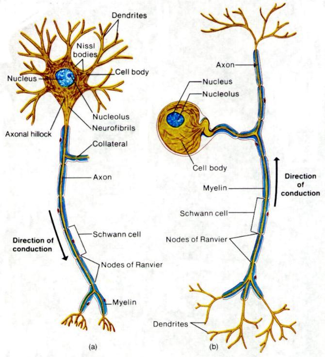
Draw a neat labelled diagram of multipolar neuron.
Neuromuscular Junction - The Definitive Guide - Biology Dictionary Web17 avr. 2021 · Even so, the synaptic gap still exists. There is no direct contact between the presynaptic and postsynaptic neuromuscular junction parts. In the electron microscope neuromuscular junction diagram below, where T indicates the motor neuron terminal and M the muscle fiber, the postsynaptic folds (arrow) are easy to see.

Neurons (Nerve Cells) Structure, Function & Types - Simply ...
how to draw structure of neuron/neuron diagram labelled ... - Pinterest Sep 13, 2020 - Please watch: "cell structure and functions / animal cell vs plant cell / parts of cell / ch 8 science class 8 cbse" ...

Common Neuron Diagram | Quizlet
Neuron - The Definitive Guide | Biology Dictionary Jun 28, 2020 · A bipolar neuron has the same structures as all neurons but is arranged differently. The central cell body makes a border between the dendrite end and the axon and terminal end. Dendrites are multiple and sometimes it is difficult to know which end is which in a simplified bipolar neuron diagram.

Diagram of Neuron Anatomy. Illustration of Neuron Anatomy ...
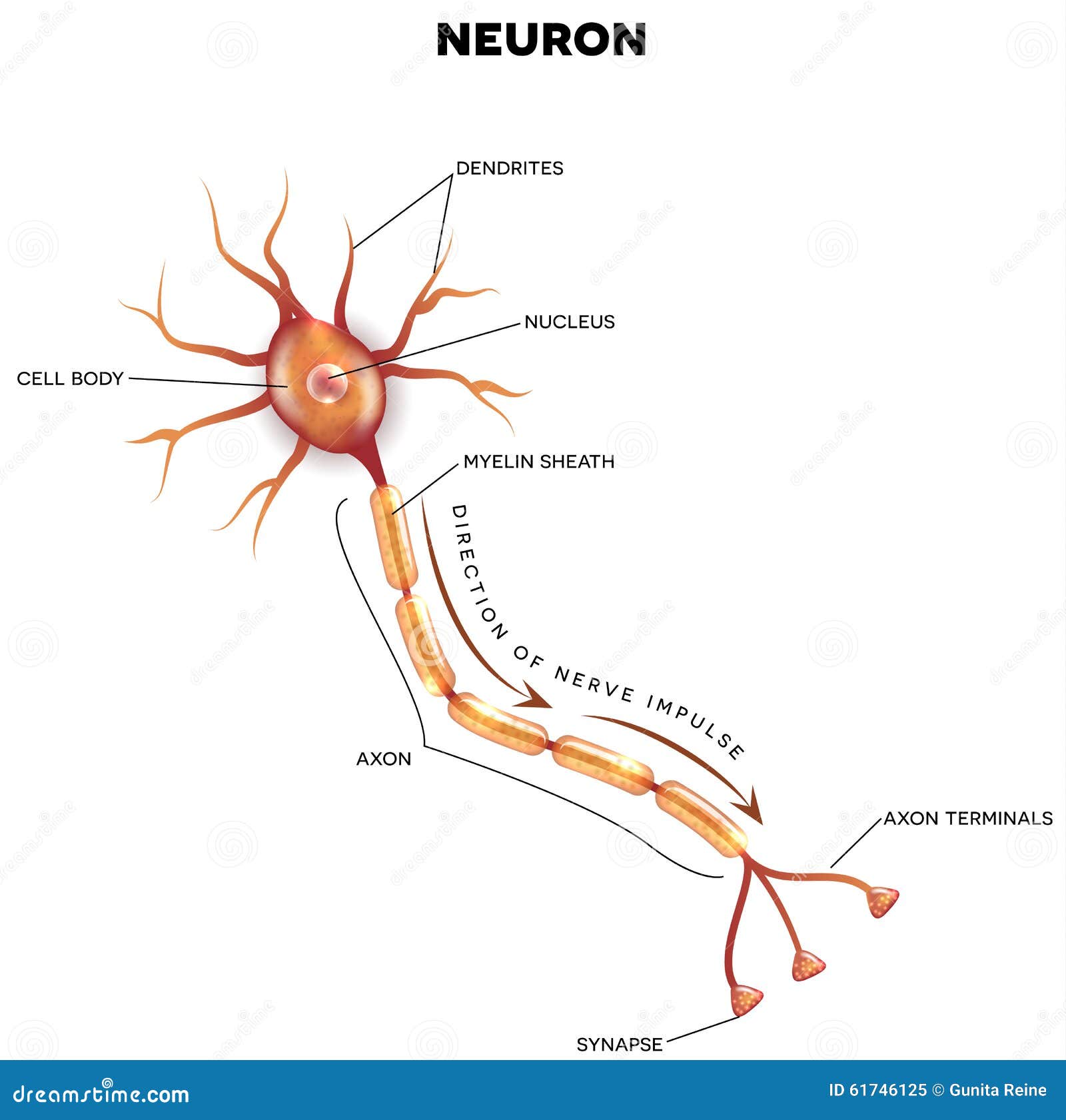
588 Neuron Labeled Images, Stock Photos & Vectors - Shutterstock Neuron that is the main part of the nervous system with description Stock Vector. Nerve Cell Neuron Labeled Diagram Stock Illustration.
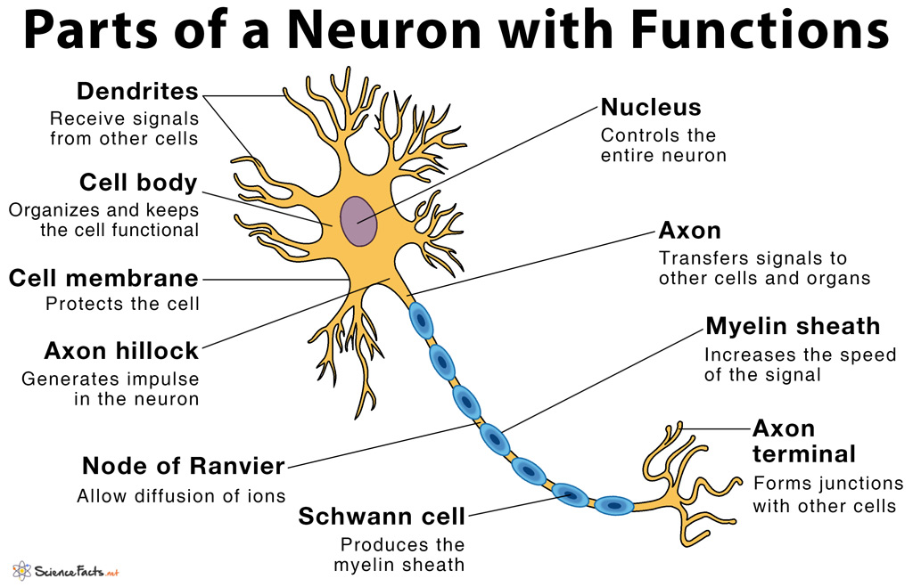
Parts of a Neuron and Their Functions with Labelled Diagram
Diagram Quiz on Neuron Structure and Function (Labeling Quiz) 2. In the figure, labeled ‘1’ receives impulses from adjacent neuron. It is called the Dendron Dendrite Axon Axonite. 3. In the figure, labeled ‘2’ is the short filaments from the cell body that carries impulses from dendrites to the cell body which is the Schwann cell Axonite Axon Dendron. 4.

Draw labelled diagrams of the following : (a) Neuron (b ...
Animal Cell- Definition, Structure, Parts, Functions, Labeled Diagram Web17 sept. 2022 · Animal cell size and shape. Animal cells come in all kinds of shapes and sizes, with their size ranging from a few millimeters to micrometers. The largest animal cell is the ostrich egg which has a 5-inch diameter, weighing about 1.2-1.4 kg and the smallest animal cells are neurons of about 100 microns in diameter.

Neuron And Synapse Labeled Diagram Royalty Free SVG, Cliparts ...
Labeled Neuron Diagram - Science Trends Types of Neurons · Unipolar: Contains only one branching body · Bipolar: Has 1 axon and 1 dendron · Multipolar: 1 axon and 2 or more dendrites · Anaxonic: Axon ...

Posts about neuron on BiologyWedsComputer | Neurons, Neuron ...
Neuron Creations - WikiEducator
CBSE Class 9 Answered
A Labelled Diagram Of Neuron with Detailed Explanations
Neurones | Anjung Sains Makmal 3

Neuron under Microscope with Labeled Diagram » AnatomyLearner ...

Describe the structure of a neuron giving a well labelled diagram

Neurons | BioNinja
How To Draw A Neuron Step By Step For Beginners

Labeled diagram of the neuron, nerve cell that is the main ...
Neuron - Wikipedia

Label Parts of a Neuron Diagram | Quizlet

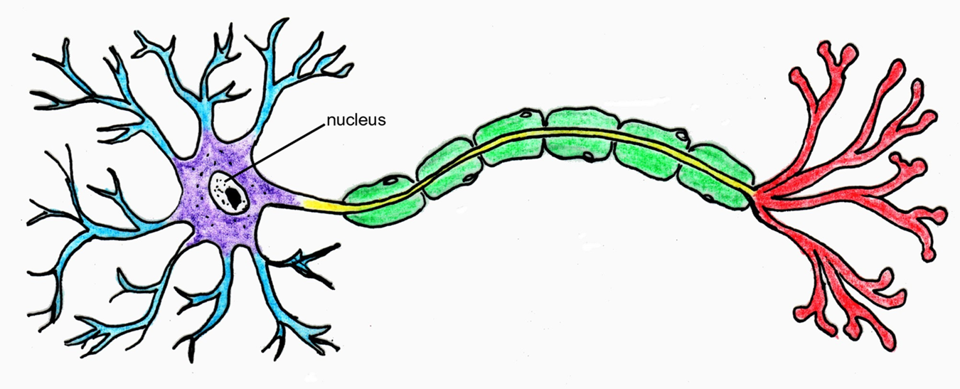
Draw a labelled diagram of a neuron.
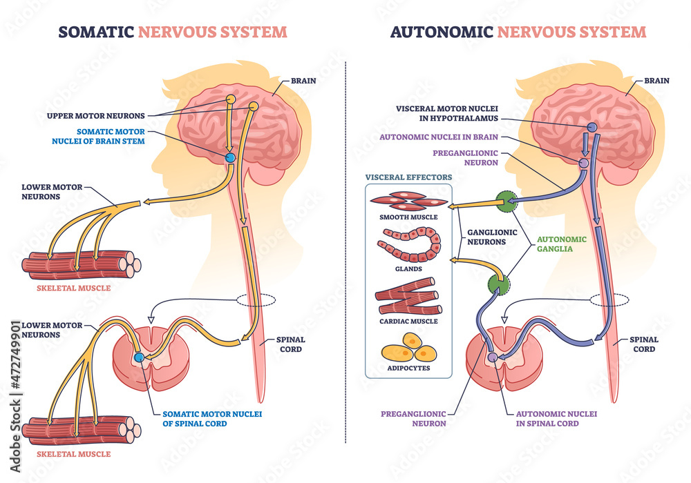
Somatic vs autonomic nervous system division in human brain ...
Types of Neurons | GetBodySmart
Labeled Neuron Diagram| EdrawMax Template

Motor neuron | Alila Medical Images
Neuron Diagram Images – Browse 2,645 Stock Photos, Vectors ...

Draw a labelled diagram of a neuron
Free Neuron Cliparts, Download Free Neuron Cliparts png ...
Lab 1 Neurohistology - Neurons
Draw a labelled diagram of a neuron
Neurons | Introduction to Psychology

Draw a labeled diagram of a nerve cell neuron

Figure, Figure showing Labeled Motor Neuron. Contributed by ...
The Neuron

Types of neurons vector illustration. labeled nerve parts ...
Labeled Diagram of the Neuron Stock Vector - Illustration of ...


Komentar
Posting Komentar